Sharper Image
Pawel Penczek and SPARX
University of Texas, Houston Medical School
Published June 4, 2012
When Pawel Penczek took his first job in the lab of Joachim Frank, a pioneer in cryo-Electron Microscopy, he had never heard about the technique. "My interest was in digital signal processing," says Penczek, now director of the Structural Biology Imaging Center at the University of Texas - Houston Medical School and lead developer of SPARX, a Cryo-EM image processing software tool. "I was only remotely aware of using EM for biological applications."
When he arrived in Frank's lab in 1989, he became part of the team working on the first cryo-EM construction of the ribosome, which finally emerged at 45A resolution. "It was a major milestone," he says. As part of the team, Penczek, who had studied physics at the University of Warsaw, took on the task of improving the image processing algorithms in SPIDER, a tool developed by the Frank lab that mathematically averages large numbers of images of a molecule to construct its meaningful 3-D representation.
Penczek, along with others in the Frank lab, spent the next 5 years on technical advancements, all in an effort to squeeze more meaning out of the data. "Only beginning from the mid-1990s did the biological payoff start," says Penczek. "That's when people started to become interested in cryo-EM findings."
Still, the effort to make cryo-EM a workable technique proved to be much more challenging than anyone had expected. "I'm still working on it 2 decades later," laughs Penczek.
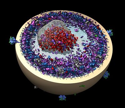
Cryo-EM's main advantage is that it allows imaging of a molecule in its native state. "Proteins appear as they are," says Penczek, "like flies in amber." In contrast, crystallized proteins used in X-ray crystallography may be constrained by the crystal.
The downside of cryo-EM is that in a single-molecule sample, the signal-to-noise ratio is very low. Solving the structure takes expertise, and, even then, the resolution is typically limited to 10 - 15 A.
For Penczek, the obvious task ahead is to improve the technique so that it becomes routine to obtain high-resolution solutions. However, says Penczek, "paradoxically, it is the resolution that I'd consider the last interesting challenge."
Rather, Penczek is focusing on finding ways to validate the resulting shape. He is also working to find ways to handle the conformational variability that comes from having unpredictable single-particle samples. "Since the molecules aren't constrained when they are frozen, they can have floppy or moving parts," he says. "We can take as many pictures as we want, but it's all fuzzy, like slow shutter speed pictures of moving cars."
According to Penczek, conformational variability is a blessing and a curse. "It limits resolution," he says, "but it also provides insight into the functioning of proteins."
For this reason, Penczek has been struggling with conformational variability for the last decade. His primary focus today is developing new image processing methods to improve validation and handle variability and implementing them in a software package called SPARX.
"SPARX is the place where my ideas take shape," he says, though his true research focus is on methods. "It's my venue for propagating ideas." For more information about SPARX, visit the SPARX Wiki at: http://sparx-em.org/sparxwiki/SparxWiki
-- Elizabeth Dougherty